• Request Info
  • Visit
  • Apply
  • Give
  • Request Info
  • Visit
  • Apply
  • Give

Search

A-Z Index Map

Physics & Astronomy

  • About
    • Honors
    • Administration
    • Faculty Resources
    • Nielsen Spaces
  • Research
    • Research Partners & Facilities
    • Condensed Matter
    • Particle / HEP
    • Biophysics / Soft Matter
    • Nuclear / Astrophysics
    • Quantum Information
  • People
    • Faculty
    • Staff
    • Joint Research & Adjunct Faculty
    • Post Docs
    • Graduate Students
  • Undergraduate
    • Why Physics
    • What Our Grads Do
    • Career Resources
    • Degree Programs
    • Research
    • Scholarships
    • Student Organizations
  • Graduate
    • Join Our Program
    • FAQs
    • Fellowships & Assistantships
    • Bains Fellowship
    • Where Our Grads Go
    • Research
    • Resources
  • News & Events
    • Newsletters
    • News
    • Colloquia Series
    • Events
    • In the Media
  • Outreach
    • Astronomy Outreach
    • Cool Things in the Sky this Month
    • Physics Outreach & High School Lecture Series
  • Alumni
    • Distinguished Alumni Award
    • Giving Opportunities
    • Share Your News
A banner showing a model of an atom and elementary particles

News

Soft Matter

A graphic showing the evolution of chromosome separation in E. coli.

UT’s Biophysics Group Investigates How Chromosomes Separate

December 16, 2025

Biologically speaking, family stories are written in chromosomes. For the story to continue, those chromosomes have to be copied and passed on to the next generation. In recently published findings, UT’s biophysicists took a deeper look at how this works in Escherichia coli (E. coli) to better understand the process.

A Simple System with a Multi-Step Process

Chromosomes are long strands of DNA that wrap around proteins. A key part of a cell’s life cycle is chromosome replication and the transfer of genetic material to daughter cells. Professor Jaan Mannik’s and Adjunct Assistant Professor Max Lavrentovich’s groups wanted to understand how that mechanism is organized and carried out in bacteria (specifically E. coli).

“Chromosomes must be equally divided between the two daughter cells during cell division, otherwise the cell that lacks a full genome will die,” Mannik explained. “In human cells, the mitotic spindle is responsible for separating the chromosomes before cell division starts. However, bacteria lack a mitotic spindle. The question arises how the bacteria separate their chromosomes.”

He explained that “it is expected, based on polymer physics models, that two new DNA strands forming during replication in cylindrical confined conditions repel from each other due to an entropic force (entropic segregation mechanism).”

The Upside of Unmet Expectations

Graduate student Chathuddasie I. Amarasinghe (a first-time first author) took on the challenge to test this mechanism experimentally. She was joined by Graduate Student Mu-Hung Chang, who tackled the same question via modeling.

A photo of Jaan Mannik
Jaan Mannik
A photo of Mu-Hung Chang
Mu-Hung Chang
A photo of Chathuddasie I. Amarasinghe
Chathuddasie I. Amarasinghe

Using high-throughput fluorescence microscopy, Amarasinghe said the group imaged thousands of cells using microfluidic devices (also called lab-on-a-chip) in a single experiment.

“We take time-lapse images of our cells and then use MATLAB and Python functions to analyze the data in different ways, both quantitatively and qualitatively, including still images and movies,” she explained.

Amarasinghe said when she first created this new strain of cells with a fluorescent tag on ribosomes, she “expected it to produce very straightforward results that would match theoretical predictions from the entropic mechanism perfectly.”

That wasn’t exactly what happened.

Working on the experiment she learned of another model proposing how the dynamics of mRNA–ribosome complexes could affect DNA segregation. Messenger RNA copies genetic material from a DNA strand and carries that information to the ribosome, which makes proteins. Amarasinghe et al. found that once the replication process is roughly at the halfway point, the accumulation of messenger RNA and ribosomes in the middle of E. coli chromosomes becomes strong enough to start driving the two daughter DNA strands away from each other. This process continues past the point when the two chromosomes lose contact with each other and separate.

In parallel to this process, Amarasinghe and co-workers found that the daughter chromosomes are also separated by the closing constriction, a final “pinching” of the cell. During constriction the cell envelope bends inward and physically pushes the chromosomes apart.

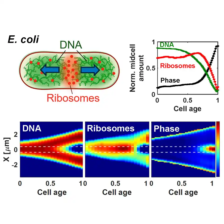
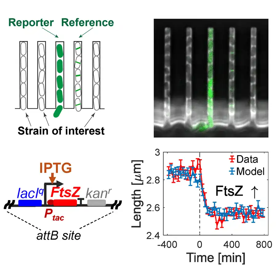
The evolution of chromosome separation in E. coli. Top left: Diagram of an E. coli cell showing polysomes pushing sister chromosomes apart.  Bottom: Heatmaps show cell-cycle-dependent changes in DNA and ribosome density distributions, and constriction formation. The top right corner shows normalized mid cell amounts from these heatmaps (integrated between the dashed horizontal lines).

Chang led development of the model, which used partial differential equations to describe the evolution of DNA, polyribosomes, and ribosomal subunits in E. coli cells.

“The model correctly predicts a local maximum in ribosome density at the cell middle in the early stages, which gives a good qualitative explanation to the DNA and ribosome correlation patterns observed in experiment,” he explained. “However, it failed to capture the exact time-scale of the accumulation of ribosomes in the cell middle in the later stages of the cell cycle. We expect this discrepancy could be potentially reduced if we extend the model to 3D in future work.” 

The results of the experiment are published in the Proceedings of the National Academy of Sciences. PNAS is a peer-reviewed journal of the National Academy of Sciences covering the biological, physical, and social sciences. In addition to Amarasinghe (first author), Chang, Lavrentovich, and Mannik, other contributors are Jaana Mannik (a research scientist with UT Physics) and Scott T. Retterer of Oak Ridge National Laboratory.

Diving into Deep Questions

Biophysics captured Chang’s attention during his first year in the physics graduate program. Lavrentovich (then on UT’s faculty) introduced his work to new students, and Chang said “the mysterious phase transition patterns occurring in living beings attracted me.”

Within a couple of years, he joined the biophysics group.

“I started studying the organization pattern of E. coli DNA, which shows phase separation behavior between different species similar to some phase transition patterns Max showed before,” he said.

Chang defended his PhD dissertation this fall and will continue working with the Mannik group while applying for postdoctoral positions.

Amarasinghe came to biophysics a little earlier, taking a biophysics course as an undergraduate.

“For my undergraduate research, I developed antimicrobial packaging materials, which gave me hands-on experience with microbiology techniques that confirmed my interest in microbial research,” she said.

When she came to UT for graduate studies and learned of Mannik’s research, she was immediately interested in becoming part of the work.

“His research focuses on understanding how life self-organizes from seemingly simple components using the model organism E. coli, which is one of the deepest open questions in biology,” she said.

December 16, 2025  |  Filed Under: Featured News, News, Soft Matter

Image from E. coli research

UT’s Biophysics Research Lays the Groundwork for Designing New Antibiotics

December 17, 2024

Experiments with E. coli cells in microfluidic channels where their length response to FtsZ levels can be determined with 50 nm precision.

Bacteria may be microscopic in size, but anyone who’s suffered through strep throat or salmonella knows they can be tough opponents. Antibiotics keep bacterial cells from dividing, stopping them in their tracks and shutting down the infections they cause. But bacteria can mutate, outsmarting these medications and making them much less effective.

To come up with new therapies that prevent bacterial cells from dividing, scientists need to know the full story of how they divide. Professor Jaan Mannik and his colleagues have made an important step in that direction by identifying which proteins are rate-limiting for cell division. The results were published in Nature Communications.

Mannik’s group studies a common strain of Escherichia coli (E. coli), which provides a good baseline for the physics of how bacteria work. He described these cells as “little rods about one-thirtieth of the diameter of a hair” and anywhere from two to four micrometers in length. In ideal growth conditions, they can divide every 20 minutes.

“Division is one of the most fundamental cellular processes,” Mannik explained. “Several well-known antibiotics, including ampicillin and cephalexin, inhibit cell division. Bacteria whose division is inhibited die after a while. If we understand division better, new antibiotics can be designed.”

There’s a pressing need for new medicines because bacteria have gained an upper hand.

“Antibiotics have been losing their effectiveness because bacteria mutate rapidly, and antibiotics are used carelessly,” Mannik said. “The latter allows the mutations to take over in cell populations. Antibiotic-resistant bacteria are not a problem of the future but a current reality.”  

Certain elements of cell division in E. coli are understood. Scientists know it begins with the formation of a ring-shaped structure (the Z-ring) around the cell’s middle. The Z-ring organizes proteins to develop a septal wall. However, the formation of the Z-ring is not yet sufficient to trigger cells to constrict—a process responsible for splitting a mother cell into two daughters.

There are a few similar-sounding proteins at work here: FtsZ (Filamenting temperature-sensitive mutant Z), as well as FtsN and FtsA. Earlier research has shown that FtsZ is critical for forming the Z-ring, but Mannik and his colleagues were looking for its role in triggering the constriction, which happens long after the Z-ring has formed.

“We want to determine what mechanism triggers their division,” he said. “In other words, how (do) a bunch of proteins, such as FtsZ and FtsN, assemble in the cell and ‘decide’ that cell needs to divide?”

To discover what flips the switch, Jaan Mannik worked with Jaana Mannik, a research scientist in the department, as well as graduate student Chathuddasie Amarasinghe and colleagues from Harvard University and the Weizmann Institute of Science who modeled the division process. He said others in the field have thought FtsN was the key protein prompting cell division in E. coli, “but our research shows that it does not.”

The team got their experimental results using high throughput imaging in microfluidic devices—essentially a miniature lab on a quarter-size chip that lets researchers grow individual bacterial cells in microscopic channels, prod them with different stimuli, and then record their responses. Mannik said their key finding is that FtsZ accumulation controls when cells start to constrict. FtsZ numbers in the cell must reach a certain threshold level to trigger the process. This threshold level allows FtsZ filaments to form doublets or bundles, which recruit some other Fts-proteins and trigger a cascade of reactions needed for cells to constrict.

“If the constriction does not start to form, cells grow to long filaments and then die,” he said. “If we understand how this process takes place in E. coli and presumably also in other bacteria, then we can devise new means to stop it and thereby treat bacterial infections.”

December 17, 2024  |  Filed Under: Featured News, News, Soft Matter

Photo of Dima Bolmatov

Where Are Your Memories Stored?

May 10, 2023

Bolmatov

Dima Bolmatov explains in The Conversation

You may easily remember your kindergarten teacher, your brother’s birthday, or how to do long division. But how? Why do these things get filed away in a place you can quickly access while other information gets lost? Research Assistant Professor Dima Bolmatov helps explain the biophysics of memory for The Conversation in Memories May be Stored in the Membranes of your Neurons.

The article takes findings reported in the Proceedings of the National Academy of Sciences and reframes the science for a general audience. The original research concerns the physics of the brain’s infrastructure and how it influences learning and memory. Understanding how our brains encode information for retrieval has implications for both medicine (Alzheimer’s and dementia research) and computing (developing the neural networks that drive machine learning).

Bolmatov joined the physics department in 2020 and works with the Shull-Wollan Center (SWC) at Oak Ridge National Laboratory. The center connects him with the requisite tools (e.g., X-ray and neutron scattering) to study biological systems at the molecular level. His research links biophysics and soft condensed matter and he collaborates with Assistant Professor Max Lavrentovich and Professor Alan Tennant as well as colleagues in biology, psychology, and materials science.

May 10, 2023  |  Filed Under: Featured News, News, Soft Matter

Recent Posts

  • Physics After Dark
  • How Spin Shapes the World
  • Cosmic Collaboration: Students Join Forces to Bring the Invisible to Life
  • UT’s Biophysics Group Investigates How Chromosomes Separate
  • WiP Closes Fall Term with Lunch & Networking

Physics & Astronomy

College of Arts and Sciences

401 Nielsen Physics Building
1408 Circle Drive
Knoxville TN 37996-1200
Phone: 865-974-3342
Email: physics@utk.edu

Facebook Icon    X Icon

The University of Tennessee, Knoxville
Knoxville, Tennessee 37996
865-974-1000

The flagship campus of the University of Tennessee System and partner in the Tennessee Transfer Pathway.

ADA Privacy Safety Title IX