UT’s Biophysics Research Lays the Groundwork for Designing New Antibiotics
Bacteria may be microscopic in size, but anyone who’s suffered through strep throat or salmonella knows they can be tough opponents. Antibiotics keep bacterial cells from dividing, stopping them in their tracks and shutting down the infections they cause. But bacteria can mutate, outsmarting these medications and making them much less effective.
To come up with new therapies that prevent bacterial cells from dividing, scientists need to know the full story of how they divide. Professor Jaan Mannik and his colleagues have made an important step in that direction by identifying which proteins are rate-limiting for cell division. The results were published in Nature Communications.
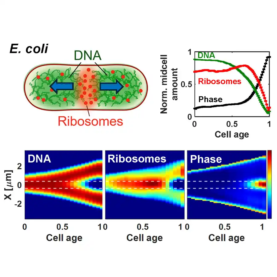
Mannik’s group studies a common strain of Escherichia coli (E. coli), which provides a good baseline for the physics of how bacteria work. He described these cells as “little rods about one-thirtieth of the diameter of a hair” and anywhere from two to four micrometers in length. In ideal growth conditions, they can divide every 20 minutes.
“Division is one of the most fundamental cellular processes,” Mannik explained. “Several well-known antibiotics, including ampicillin and cephalexin, inhibit cell division. Bacteria whose division is inhibited die after a while. If we understand division better, new antibiotics can be designed.”
There’s a pressing need for new medicines because bacteria have gained an upper hand.
“Antibiotics have been losing their effectiveness because bacteria mutate rapidly, and antibiotics are used carelessly,” Mannik said. “The latter allows the mutations to take over in cell populations. Antibiotic-resistant bacteria are not a problem of the future but a current reality.”
Certain elements of cell division in E. coli are understood. Scientists know it begins with the formation of a ring-shaped structure (the Z-ring) around the cell’s middle. The Z-ring organizes proteins to develop a septal wall. However, the formation of the Z-ring is not yet sufficient to trigger cells to constrict—a process responsible for splitting a mother cell into two daughters.
There are a few similar-sounding proteins at work here: FtsZ (Filamenting temperature-sensitive mutant Z), as well as FtsN and FtsA. Earlier research has shown that FtsZ is critical for forming the Z-ring, but Mannik and his colleagues were looking for its role in triggering the constriction, which happens long after the Z-ring has formed.
“We want to determine what mechanism triggers their division,” he said. “In other words, how (do) a bunch of proteins, such as FtsZ and FtsN, assemble in the cell and ‘decide’ that cell needs to divide?”
To discover what flips the switch, Jaan Mannik worked with Jaana Mannik, a research scientist in the department, as well as graduate student Chathuddasie Amarasinghe and colleagues from Harvard University and the Weizmann Institute of Science who modeled the division process. He said others in the field have thought FtsN was the key protein prompting cell division in E. coli, “but our research shows that it does not.”
The team got their experimental results using high throughput imaging in microfluidic devices—essentially a miniature lab on a quarter-size chip that lets researchers grow individual bacterial cells in microscopic channels, prod them with different stimuli, and then record their responses. Mannik said their key finding is that FtsZ accumulation controls when cells start to constrict. FtsZ numbers in the cell must reach a certain threshold level to trigger the process. This threshold level allows FtsZ filaments to form doublets or bundles, which recruit some other Fts-proteins and trigger a cascade of reactions needed for cells to constrict.
“If the constriction does not start to form, cells grow to long filaments and then die,” he said. “If we understand how this process takes place in E. coli and presumably also in other bacteria, then we can devise new means to stop it and thereby treat bacterial infections.”